Integrative Medicine Case Reports, Volume 4, Issue 2 (July), 2023
Bone marrow mesenchymal stem cells and exercise restore motor function following spinal cord injury by activating PI3K/AKT/mTOR pathway. Neural Regen Res. 2023 May;18(5)1067–1075
ABSTRACT |
||
*Corresponding Author:
|
Despite the fact that a number of therapeutic modalities have shown promise in the treatment of spinal cord injury (SCI), focusing solely on one aspect of recovery will not lead to successful and useful regeneration in people. The current study targets an integrated approach of neuroprotection and rehabilitation strategies to treat spinal cord injury, increasing nerve regeneration and functional recovery through functional sensorimotor training and cell transplantation. Sun et al., 2022 investigated the synergistic effect of exercise training and bone marrow mesenchymal stem cell transplantation which could promote active recovery in a mice model of thoracic contusive spinal cord injury. doi: 10.38205/imcr.040277 |
Introduction
Spinal cord injury (SCI) is a life-altering neurological condition for which no possible treatments have yet been identified. SCI is caused by trauma, inflammatory disorders, tumours, vascular anomalies, and gradually degenerative cervical myelopathy (1).
SCI pathophysiology is divided into two categories: primary and secondary damage. Compression, shearing, stretching, and traction forces can all cause initial mechanical damage. Damage to the central and peripheral nervous systems is caused by direct compression of neural tissue by bone fragments, disc debris, and ligaments. As a result, blood vessels are harmed, axons are disturbed, and brain cell membranes are ruptured. Microhaemorrhages occur in the core grey matter within minutes and grow radially and axially during the next few hours. The primary injury is followed by a series of subsequent events that aggravate neurological dysfunction and expand the extent of tissue harm (2).
So far, numerous neurogenerative treatments for SCI have been investigated, and stem cell transplantation is one of the prospective therapy for SCI. According to previous studies, administration of bone marrow mesenchymal stem cells (BM-MSCs) in SCI model can be a effective therapy as they enhance tissue repair and functional recovery (3,4). In addition, Massoto et al., 2020 have shown that combination therapy of mesenchymal stem cells and exercise promotes tissue repair better than stem cells or exercise individually (5).
Hence, current study targets at synergistic therapy of BM-MSCs and exercise to treat SCI in mouse model (Figure 1).
Figure 1: Schematic representation of combinatorial therapy of BMMSCs and TMT in SCI mice model. (Created with BioRender.com.)
Study Design
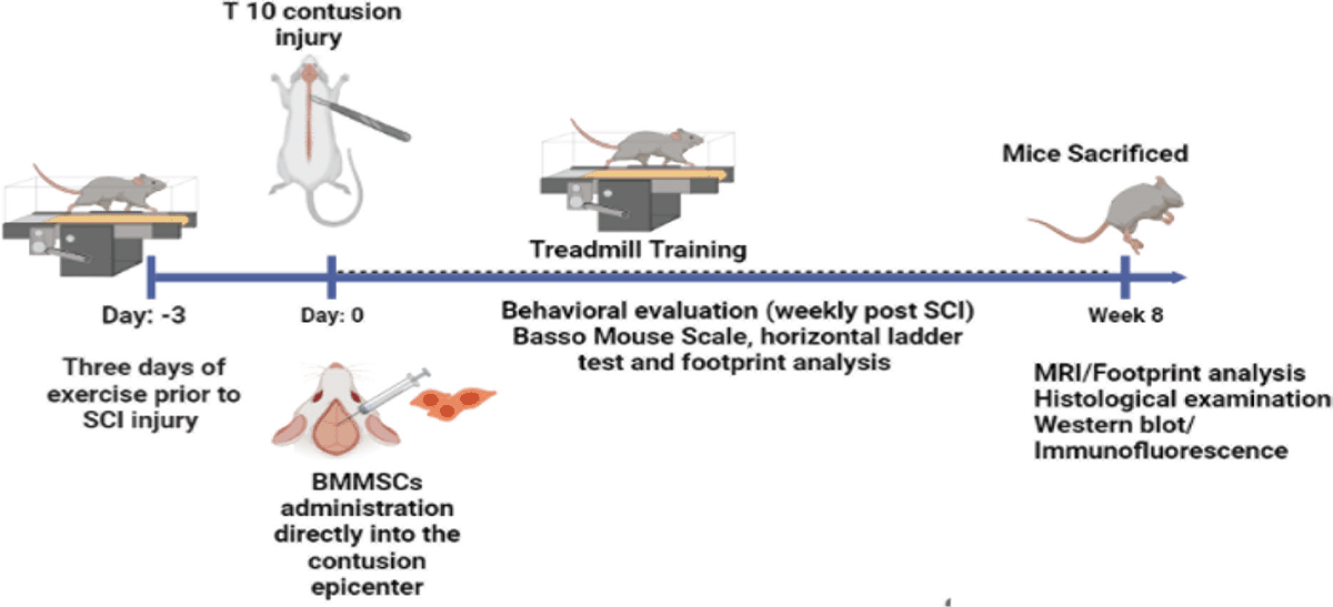
All the experiments had due approvals from Laboratory Animals Ethics Committee of the West China Hospital of Sichaun University. C57BL/6 female mice (n = 83) were procured from Chengdu Dossy Experimental Animals Co., Ltd. Among 83 mice, 8 mice were being used in the procedure for BM-MSCs isolation and remaining 75 mice were used for SCI studies. Before generating SCI model, mice in TMT and BM-MSCs+ TMT group were given treadmill training (TMT) on treadmill apparatus at speed 4 m/min to the maximum 9 m/min (Figure 2). Treadmill training was given for 8 weeks (6 days/week for 20 minutes).
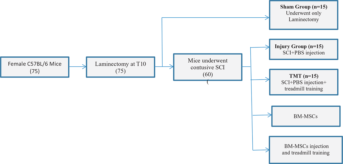
Figure 2: Schematic summary of experimental groups.
Various methods were used to study the effect of BM-MSCs and TMT in different groups of SCI.
1. Behavioral evaluation
2. Transmission electron microscopy
3. Histological examination
4. Magnetic resonance imaging
5. Western blot analysis
6. Immunofluorescence
Results
1. Synergistic treatment of mice with BMMSCs and TMT has enhanced the locomotor recovery. For behavioral studies, BMS (Basso Mouse Scale) score assessment, foot print analyses and horizontal ladder test were done. As a result, BMS score was higher in BM-MSCs and TMT treated group as compare to injury group (Figure 3). Similarly, in footprint and horizontal ladder test, mice treated with BM-MSCs and TMT showed better locomotor movement and a significant decrease in error rates; respectively.
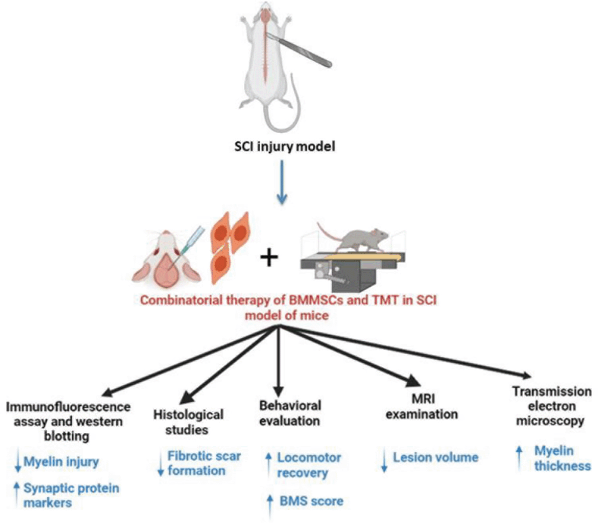
Figure 3: Schematic representation of effect of combinatorial therapy in SCI model of mice. (Created with BioRender.com.)
2. T2-weighted MRI examination proved that there was significant decrease in lesion volume in mice administered with BM-MSCs and TMT. Also, histological studies has revealed that combined treatment has significantly affected the fibrotic area and has lower the fibrotic scar formation.
3. Combinatorial treatment of BM-MSCs and TMT has neuroprotective effect in SCI mice (Figure 3). It was demonstrated by double immunofluorescent staining for GFAP (Glial fibrilliary acidic protein) and NeuN (Neuronal Nuclear Antigen) and it showed high NeuN density. Hence, high neuronal density in BM-MSCs+ TMT group as compared to other groups was present. Further, Nissl staining showed that only BM-MSCs+ TMT group exhibited a significant increase in the number of surviving neurons as compared to the injury group. Therefore, it promoted repair of damaged tissue.
4. Double immunofluorescent staining for GFAP and NF200 (an axonal marker) indicate that the combination therapy can alleviate axon and myelin destruction in the injured site compared to TMT or BMMSC alone. Further, transmission electron microscopy revealed that combinatorial therapy promotes remyelination, hence increases the myelin thickness (Figure 3).
5. To evaluate the synaptic function, immunofluorescence staining of PSD (a presynaptic marker protein) and SYN (a post synaptic marker protein) was performed followed by western blotting. It indicated that combination therapy upregulate the expression of synaptic protein markers and improves the synaptic function (Figure 3).
Implication
This study reveals that BM-MSC transplantation combined with exercise training synergistically improved locomotor activity in paralyzed hindlimbs and promotes functional restoration. Combination therapy promotes axon and myelin protection, enhances synaptic function and neurotrophin secretion, suppressing scar formation. In addition, neuroprotective effect of BM-MSCs and TMT is mediated through upregulation of PI3K/Akt/mTOR pathway. It was shown by blocking PI3K/AKT/mTOR signaling pathway in vitro in PC12 (rat pheochromocytoma) cell line and rat primary cortical neurons, which exacerbates neuronal injury.
Received Date: 12-06-23; Revised Date: 27-06-23
Accepted Date: 28-06-23
References
1. Guest J, Datta N, Jimsheleishvili G, Gater DR. Pathophysiology, Classification and Comorbidities after Traumatic Spinal Cord Injury. J Pers Med. 2022;12(7).
2. Siddall PJ, Wrigley PJ. Spinal cord injury. Clin Pain Manag Chronic Pain, Second Ed. 2008;359:388–404.
3. Lin L, Lin H, Bai S, Zheng L, Zhang X. Bone marrow mesenchymal stem cells (BMSCs) improved functional recovery of spinal cord injury partly by promoting axonal regeneration. Neurochem Int [Internet]. 2018;115:80–4. Available from: https://doi.org/10.1016/j.neuint.2018.02.007
4. Liu NK, Xu XM. Neuroprotection and its molecular mechanism following spinal cord injury. Neural Regen Res. 2012;7(26):2051–62.
5. Massoto TB, Santos ACR, Ramalho BS, Almeida FM, Martinez AMB, Marques SA. Mesenchymal stem cells and treadmill training enhance function and promote tissue preservation after spinal cord injury. Brain Res [Internet]. 2020;1726(September 2019):146494. Available from: https://doi.org/10.1016/j.brainres.2019.146494