Integrative Medicine Case Reports, Volume 5, Issue 1 (January), 2024
Plausible role of homoeopathic intervention in the management of Tinea Faciei; A case series
KEY WORDS |
ABSTRACT |
|
Dermatophytosis
|
Around the globe, dermatophyte infections are an increasingly prevalent type of superficial fungal infection. Several dermatophytes have a varying global distribution, where few are confined themselves to a region in particular. The nosological name of superficial dermatophyte infection varies according the location of the body, but in particular the variant that affects skin of the face is known as tinea faciei. It is frequently clinically misdiagnosed due to its variable appearance. The lancet reported it as an epidemic of fungal infection and nowadays occurrence has been fairly common OPDs and in clinical practice. Despite of being common in occurrence dermatophytosis is still a neglected topic in medical research. Different level of evidences like caser-report, case-series and RCT has been published on dermatophytosis but till date no evidence has been reported on tinea faciei only. Apart from quality of life tinea faciei has a significant impact as it can cause cosmetic disfigurement due to it’s site. Here we are reporting about five cases of tinea faciei which has been managed efficiently with the help of individualized homeopathic medicine. Natrum muriaticum and Nux vomica was commonly indicated in the cases what we have treated. In all the cases significant improvement was seen without any further complications. In three out of five cases the patient was previously treated with anti-fungal combinations without much benefit, which depicts why homeopathy should be considered in the possible integration in the treatment of such fungal diseases. Though we couldn’t specify the strains because of not having proper infrastructure but that is not essential for the successful homeopathic prescription. In future more structure and well designed clinical trial is desired to understand the scope and limitations for better clinical application.
doi: 10.38205/imcr.050118 |
|
*Corresponding Author:
|
Introduction
The skin of humans is a multilayered ecosystem with a wide range of micro-environmental factors, including incredibly varied and intricate communities of microbes (1). Although the history of superficial cutaneous infections is lengthy, Chester Emmons initially categorized dermatophytes in 1934 into the following three genera: Trichophyton, Microsporum, and Epidermophyton (2). This was based on the morphology of the spores and the accessory organs. Subsequently, nine distinct species were found, with the four genera Trichophyton, Microsporum, Epidermophyton, and Nannizzia containing the majority of human diseases (3,4). Over time, the organism causing dermatophytosis has evolved. Microsporum audouinii and Trichophyton schoenleinii were the most common species in the early 20th century, whereas T. rubrum and T. mentagrophytes took over in the late 20th century (5–7). In the 21st century along with the increasing burden of T. rubrum, there has been a concerning rise in the prevalence of antifungal-resistant species within certain ethnic groups, further complicating the management of these infections (8). A multicenter study conducted in India, involving clinically diagnosed dermatophytosis patients and molecular analysis of 351 specimens, reported that T. indoline was the most prevalent species (90%), followed by T. rubrum (5%) and T. mentagrophytes/T. interdigitale (5%) (9). T. indoline has demonstrated reduced susceptibility to azole drugs and resistance to terbinafine, with over 50% of the strains exhibiting resistance (9,10). Reports of T. indoline have also emerged in the Middle East (Iran), Asia, and Europe in recent years (11). Unlike other parts, fungal infections of the face are not common in routine dermatological practice. Facial skin infections can occur from external sources (e.g. infected pets or other people) or from direct inoculation of a fungus after infection at a remote site in the affected patient (“self-inoculation”) (12,13). Tinea faciei may simultaneously occur secondary to other forms of tinea infection, particularly tinea capitis and tinea corporis, or rarely face becomes the primary site of infection. There is an increasing prevalence of cutaneous dermatophytosis across the world, and especially in the tropics and India is not an exception to that (14,15). In India alone, the prevalence ranges from 36.6–78.4% which could be attributed to tropical climate and lack of awareness on personal hygiene (16). The change in prevalence of dermatophytes causing the disease is the result of host factors such as an increase in cases of co-morbidity and immune suppression; hot and humid tropical climate, lifestyle changes, and poor hygiene etc. (17). Though several studies had been conducted in different states of this country unfortunately no studies worth mentioning have been found from West Bengal (18). In general fungal infections are mostly neglected area of research but the present scenario promptly calls for more research work in this area. Recent studies show a positive family history of dermatophytosis as a major risk factor along with the previously established relationship with Diabetes Mellitus (19). These lesions have a wide range of clinical presentations; tinea cruris and tinea faciei are the most frequent. Additionally, the clinical behavior of ringworms has been steadily changing. This includes the ability to spread quickly to form larger lesions, to involve distant regions early on, to easily spread to other family members, to flare up symptoms after treatment with occasional systemization, to coexist with bacterial infections, and to relapse chronically (8). Drug-resistant strains are posing new issues due to their shift in character, which can be attributed to the intricate interactions between host environment and agent variables. The increased irrational misuses of unwanted corticosteroids, anti-fungals, anti-bacterial combinations, that are cheap and easily available over the counter, have increased in resistant and non-responsive lesions, which increase the disease burden on the society (17). Combined with the poor quality of life, the long-term cost of treatment in the conventional system of medicine further adds to the plight of the patient. Homeopathy is a popular choice of treatment following modern medicine having justifiable scope in the treatment of such cases in a far more cost-effective way without leaving behind further complications (20). Previously we have published a case series of dermatophytosis depicting the scope of homeopathy (21). Here in this case report we are willing to depict the scope of individualized homeopathy in the management of Tinea faciei.
Case Report-I
Patient Information
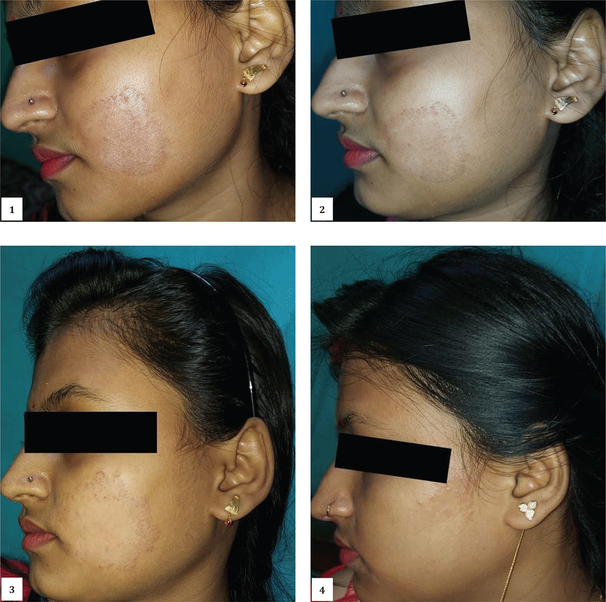
A female patient aged about 19 years, accompanied by her mother, visited the clinic with complaints of a large circular area of itching and eruption on her left cheek [Figure 1]. She had been suffering from the condition for the last year. She had opted for allopathic medicine in the beginning with a temporary relief of 3 months, but the lesions reappeared, and since then it have been resistant to treatment. During case-taking, it was noted that she had a good appetite with a strong desire for meat and salty food. She had a profuse thirst and a dry, white-coated tongue. She has tended to catch colds easily, since childhood. All her complaints aggravated in the summer months. She had a fear of downward motion and ghosts. She desired company at all times and preferred conversation.
Clinical Findings
An erythematous patch with a distinct margin is present on the left cheek. The area was hot to the touch. The itching aggravated in the evening and was ameliorated by continued scratching. There was no discharge from the lesion. Careful clinical examination led us to diagnose the condition as Tinea faciei. As the prescription does not depend on the subtype of the dermatophytes we didn’t go for a KOH test or culture.
Therapeutic Intervention
Based on the totality of symptoms 2 doses of Borax 30CH were prescribed. The patient was called for follow-up after 3 weeks. Gradual improvement was seen. Borax 30/2 doses were again prescribed on 17-02-2022. She was advised to visit after 5 weeks [Figures 2–4]. The next follow-up was taken on 24-03-2022, based on the symptoms during the follow up Natrum Mur 1M/2 doses were given (Table 1). The patient showed gradual and consistent improvement thereafter. She was given a placebo on follow-up.
Figures 1–4: Showing chronological development and remission of the lesion during the course of treatment of case-I.
Table 1: Showing Prescriptions and Time line of Case-1
| Date | Symptoms | Medicines Prescribed |
|---|---|---|
| 20-01-2022 | Dry itching eruption on left cheek, hot to touch, itching< evening, > scratching | Borax 30/2D |
| 03-02-2022 | Itching reduced | Placebo |
| 17-02-2022 | No itching present, lesion same as before | Borax 30/2D |
| 24-03-2022 | Standstill condition | Natrum Mur 1M/2D |
| 21-04-2022 | Reduced size of lesion with no itching | Placebo |
| 19-05-2022 | Reduced mark of lesion with no itching | Placebo |
| 23-06-2022 | No mark present with no itching | Placebo |
Follow-up and Outcome
During subsequent follow-ups, the potency and medicine were changed based on the assessment of the improvement of the lesion [Table 1]. Finally, the lesion of tinea disappeared gradually. Reappearance of symptoms was not reported even after 9 months of treatment for other complaints. A detailed timeline of treatment is mentioned in Table -1 and an analysis of MONARCH score is given in Table 7.
Case Report-II
Patient Information
A 50-year-old lady came to the clinic accompanied by her sister on the 13th of April, 2022, with the complaint of a circular itching spot on the left temporal region, near the eye. She has been suffering from the condition for the last 8 months [Figure 5]. In the beginning, she had used over-the-counter ointments, and the symptoms reduced for a couple of weeks thereafter, but, soon the symptoms flared up. The eruptions cause problems in her day-to-day life now. No medical advice was sought before this visit. Detail history taking revealed that her mother is suffering from essential hypertension for last 7 years. She has undergone a total hysterectomy following complaints of profuse painful menses about a year ago. The eruptions began a few months after the hysterectomy.
Clinical Findings
An erythematous circular patch with a distinct margin is present on the left temporal region, near the eye. Severe itching is present which aggravates while outside in the sun, sweating, or in heat. The itching reduces on applying cold to the lesion. There is no discharge from the lesion. On further case-taking, it was noted that she had very little or no appetite at all and could go without meals for long hours. She had a profuse thirst and a slightly white-coated moist tongue. She had increased urgency for urination, mostly during the day. She was reported to be angered easily and did not prefer to be consoled by others. She preferred the company of people and meeting new people.
Therapeutic Intervention
Considering her age and a few of her symptoms she was advised to investigate blood for FBS and PPBS. Based on the totality of symptoms, Natrum Mur 200/2 doses were prescribed. She was asked to report after 2 weeks. On the next visit, the blood reports were within the normal range. Her symptoms had improved. The itching had reduced. She was prescribed Placebo and advised to report after 1 month. After 1 month the lesion had almost disappeared. She was prescribed Placebo for 1 month and advised to report after 1 month. After 1 month there was no sign of the lesion [Figure 6].
Follow-up and Outcome
As the patient showed steady improvement following the first prescription, she was prescribed Placebo in all her consecutive visits. The lesion finally resolved and was not reported to reappear after 6 months of treatment. A detailed timeline of treatment is mentioned in Table 2 and an analysis of MONARCH score is given in Table 8.
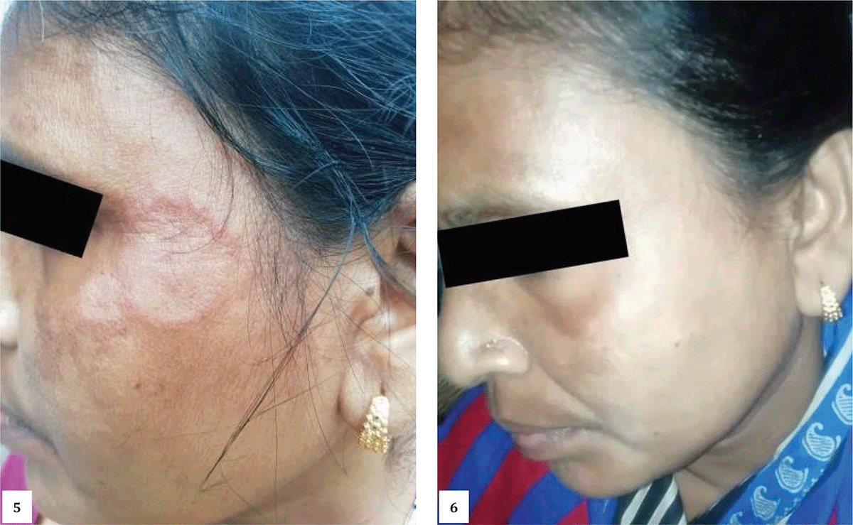
Figures 5–6: Showing chronological development and remission of the lesion during the course of treatment of case-II; here we have seen marked improvement was noted in the second visit.
Table 2: Showing Prescriptions and Time line of Case-2
| Date of visit | Symptoms | Medicines Prescribed |
|---|---|---|
| 13-04-2022 | Dry itching eruption on left temporal region, near the eye, itching < in the sun, on sweating, in heat, > on cold application | Natrum Mur 200/2D |
| 27-04-2022 | Itching reduced | Placebo |
| 25-05-2022 | No itching present, lesion reduced | Placebo |
| 29-06-2022 | Reduced size of lesion with no itching | Placebo |
| 27-07-2022 | No lesion seen | Placebo |
Case Report-III
Patient’s information
A male patient PP aged about 36 years came to our OPD with complaints of itching and eruption in the face for the last 6 months [Figure 7]. He has consulted a dermatologist and has been using anti-fungal creams for the last 6 months. During the initial course of treatment, the patient was improving but in the last 2 months his complaints have increased and the area is gradually increasing. The patient is better only while consuming anti-allergic medicines; otherwise, the severity of the itching is gradually increasing every single day. Complaints aggravate mostly during the daytime without any significant alleviating modalities.
Clinical Finding
During examination, a circular lesion was seen on the right cheek of the face without any discharge. Mild hyper-pigmentation was noted which was possibly due to the application of ointment. Systemic examination revealed an old scar of tinea cruris which was probably treated with the application of steroids. On detailed case taking the patient revealed that the itching was getting aggravated from washing at night. The patient further revealed that he had chicken pox at 12 years of age and viral hepatitis at the age of 27 years. Detailed family history failed to give any positive history of skin disease among the parents and brothers. A general investigation of the patient reveals his appetite is increased in the evening and has a desire for sour things and meat with a dislike for sweet things; the patient also prefers to eat warm food. The patient has moderate thirst with a clean moist tongue having a red tip. The perspiration is profuse in the face and head with an offensive smell and urine is within normal limits. The patient has occasional constipation and has to go for stool twice daily. Sleep is undisturbed but anxious about his health when awake. Hot patient and irritable at trivial ailments. Symptoms were repertorized for this case and the repertorization table is available in Table 3.
Table 3: Showing Repertorization table of Case-3
| Remedy | Sulph | Ars | Nit-ac | Sep | Phos | Lyc | Ferr | Puls | Lach | Graph | Nat-m | Sil | Arg-n | Arn | Fl-ac |
|---|---|---|---|---|---|---|---|---|---|---|---|---|---|---|---|
| Totality | 18 | 15 | 14 | 13 | 12 | 11 | 10 | 10 | 10 | 10 | 9 | 9 | 8 | 8 | 8 |
| SymptomsCovered | 9 | 7 | 6 | 6 | 6 | 5 | 6 | 6 | 5 | 4 | 5 | 5 | 5 | 5 | 5 |
| [Kent] [Face] Itching (seeskin): Cheeks: | 1 | 0 | 0 | 0 | 0 | 0 | 0 | 1 | 0 | 0 | 1 | 0 | 0 | 0 | 0 |
| [Kent] [Face] Itching (seeskin): Evening: | 2 | 0 | 0 | 0 | 0 | 0 | 0 | 0 | 0 | 0 | 0 | 0 | 0 | 0 | 0 |
| [Kent ] [Stomach] Appetite: Increased (hungeringeneral): Evening: | 0 | 0 | 0 | 2 | 0 | 1 | 0 | 1 | 0 | 0 | 2 | 1 | 0 | 1 | 1 |
| [Kent ] [Stomach] Desires: Sour, acids, etc.: | 2 | 2 | 0 | 2 | 2 | 0 | 2 | 2 | 2 | 0 | 2 | 0 | 1 | 2 | 2 |
| [Kent] [Stomach] Desires: Meat: | 1 | 0 | 0 | 0 | 0 | 0 | 1 | 0 | 0 | 1 | 1 | 0 | 0 | 0 | 0 |
| [Kent ] [Stomach] Desires: Warm: Food: | 0 | 3 | 0 | 0 | 0 | 2 | 2 | 0 | 0 | 0 | 0 | 1 | 0 | 0 | 0 |
| [Kent] [Stool] Hard: | 3 | 2 | 3 | 3 | 3 | 3 | 2 | 2 | 3 | 3 | 3 | 3 | 2 | 1 | 1 |
| [Kent] [Stomach] Aversion: Sweets: | 2 | 2 | 1 | 0 | 2 | 0 | 0 | 0 | 0 | 3 | 0 | 0 | 0 | 0 | 0 |
| [Kent ] [Perspiration] Odour: Offensive: | 3 | 2 | 3 | 3 | 2 | 3 | 2 | 3 | 2 | 3 | 0 | 3 | 0 | 3 | 2 |
| [Kent ] [Mouth] Discoloration: Tongue: Red: Tip: | 3 | 3 | 2 | 0 | 0 | 2 | 1 | 0 | 2 | 0 | 0 | 0 | 3 | 0 | 2 |
| [Kent ] [Mind] Anger, irascibility (see Irritability, Quarrelsome): Trembling, with: | 0 | 0 | 2 | 1 | 1 | 0 | 0 | 0 | 0 | 0 | 0 | 0 | 1 | 0 | 0 |
| [Kent] [Mind] Anxiety: Health, about: | 1 | 1 | 3 | 2 | 2 | 0 | 0 | 1 | 1 | 0 | 0 | 1 | 1 | 1 | 0 |
Therapeutic Intervention
Sulphur 200/2D was prescribed to the patient after a review of the reportorial analysis (Table 3) and the entirety of the information. A follow-up was conducted every month. In the follow-up visits that followed, a placebo was administered in light of the patient’s progressive improvement [Figures 8–10]. Table 4 provides a comprehensive timeline of the treatment, while Table 9 presents an examination of MONARCH.
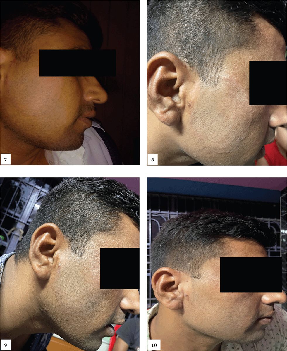
Figures 7-10: Showing chronological development and remission of the lesion during the course of treatment of case-III.
Table 4: Showing Prescriptions and Time line of Case-3
| Timeline | Symptoms | Prescriptions |
|---|---|---|
| 05/03/2023 | Itching eruptions of right cheeks aggravated at night and washing. Desire for meats and warm foods, aversion to sweets. Offensive perspirations, red tip tongue.easily angered with trembling of limbs. | 1. Sulphur 200/2 D, OD × 2 days
2. Rubrum 30/28 Doses × OD |
| 08/05/2023 | Itching slightly reduced than before but, eruptions still persist. | Placebo |
| 11/06/2023 | Itching reduced, eruptions also slightly reduced. | Placebo |
| 17/07/2023 | Itching and eruptions both reduced and the patient is better than before. | Placebo |
Case Report-IV
Patient’s Information
A 46-year-old female patient SM visited the OPD with complaints of itching and rashes for the last 6 months [Figure 11]. Previously the patient was under the supervision of a dermatologist who prescribed her anti-fungal creams for topical application and anti-allergic drugs to combat the itching. After careful examination, it was revealed that the patient had a similar kind of lesion in the groin as well. On detailed history taking the patient revealed that she has been taking medicine for hypothyroidism and Type 2 Diabetes mellitus. The general information revealed that she has itching more when getting exposed to sunlight, and she feels better from the application of cold water. Dietary habits are regular non-veg with a preference for fish and salty food. Stool and urine were within normal limits but the patient had excessive sweating over the head and all over her face. Mentally alert, quick, and active patient but she feels irritable at trifles with an aversion to consolation.
Clinical Finding
On careful examination red erythematous lesion with scratch marks was noted all over the face. The patient was already diagnosed with Tinea faciei which led us to believe the recurrence of the same as the signs and symptoms were corresponding.
Therapeutic Intervention
After considering the symptoms the patient was prescribed 2 doses of Natrum muriaticum 200 CH followed by a placebo and asked to come after 1 month [Figures 12–13]. During the first follow-up, we found significant improvement so the patient was prescribed a placebo for 1 month. During the second follow-up patient showed marked improvement and hyperpigmentation reduced markedly. During 3rd follow up the patient’s normal appearance of the facial skin was noted and the patient was advised not to take any medicine further and to date no recurrence has been reported. A detailed timeline of treatment is mentioned in Table 5 and analysis of MONARCH score is given in Table 10.
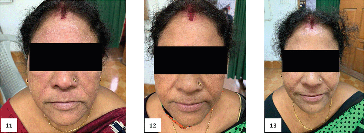
Figures 11–13: Showing chronological development and remission of the lesion during the course of treatment of case-IV.
Table 5: Showing Prescriptions and Time line of Case-4
| Date | Symptoms | Medicine |
|---|---|---|
| 1/4/2021 | Erythematous rashes with itching and scaling better from cold application. Patient has sweating over whole face with aggravates itching. | Natrum muriaticum 200/4D |
| 3/5/2021 | Itching was present to some extent and rashes were still there, but the sweating of face as reduced. | Natrum Muriaticum 200/1D |
| 14/6/2021 | Face was completely clear from and visible rashes and the patient didn’t complaint about any history of Itching. | Placebo. |
Case Report-V
Patient’s Information
In this case, a middle-aged adult female who was already undergoing treatment for PCOD and umbilical hernia came to the clinic with complaints of itching and rashes over the face [Figure 14]. On further inquiry, it was revealed that the rash has developed over the period of 2 weeks. On detailed case taking patient revealed pain on the right side of the back & and abdomen, and tenderness of the back, and that was relieved by gentle movements and during lying. Skin eruption on the left side of the face has a burning sensation which was photosensitive to the rays of sun, the itching was somewhat relieved by the use of cold water. During the period of her suffering the patient had already applied some topical combinations which she bought from an allopathic medicine shop. Further investigation revealed that she has migraine for a few months which is triggered by walking, and loud speaking. The left sided temporal headache subsides by the occurrence of vomiting. General investigation reveals she has sour eructation and a sour mouth, along with urge and stress incontinence. Both the stool and urine were very offensive in nature. The patient was unable to tolerate her hunger; she prefers to drink a small amount of water while eating, with less thirst for water. An inquiry regarding her menstrual function revealed that her LMP was 30.12.21 with regular periodical menstrual flow and occasional abdominal pain.
Clinical Finding
The circumscribed annular rash associated with central clearance and peripheral scarring has led us to diagnose the case as Tinea Faciei.
Therapeutic Intervention
After detailed case-taking, the patient was given 4 doses (D) of Nux Vomica 200 CH followed by a placebo. Again Nux Vomica 200, 2D was given after 2 months which led to complete remission of the lesion. The patient therefore continued treatment for her other ailments [Figures 15–16]. A detailed timeline of treatment is mentioned in Table 6 and analysis of MONARCH score is given in Table 11.
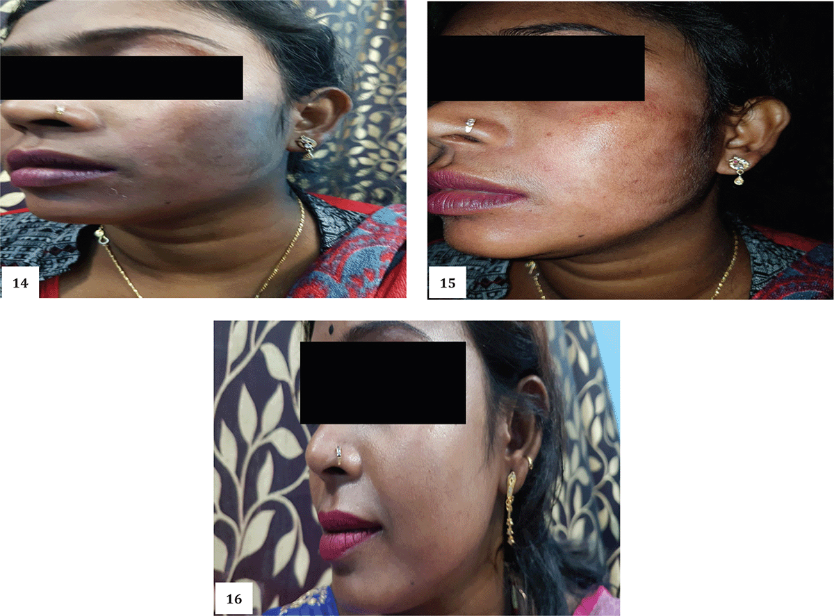
Figures 14–16: Showing chronological development and remission of the lesion during the course of treatment of case-V.
Table 6: Showing Prescriptions and Time line of Case-5
| Date | Prescription | Dose | Points Regarding Prescription |
|---|---|---|---|
| 27.2.22 | NUX. Vomica 200 | 4 | Skin eruption treated with multiple medicine for long time,
Sour eructation & mouth, irregular food habit, short tempered, industrious, headache > by vomiting, fastidious, umbilial hernia |
| 06.4.22 | Nux. Vom 200 | 2 | Sourness present, eruption persist,umbilicalherna, |
| 01.5.22 | Placebo | ||
| 25.5.22 | Merc. Sol 30 | 4 | No facial circulation,Cramping pain before stool, slimy stool, take time for stool, can not control stool with offensive, thirst profuse but saliva secration |
| 10.6.22 | Cal. Flor 200 | 4 | Back painincreased,> by gentel motion hot application,< 1st motion night, |
Table 7: Showing MONARCH score of Case-I
| Table 1(a): Modified Naranjo Criteria (MONARCH) Score | |||
|---|---|---|---|
| Items | Yes | No | Not Sure |
| 1. Was there an improvement in the main symptom or condition, for which the homeopathic medicine was prescribed? | +2 | ||
| 2. Did the clinical improvement occur within a plausible time frame relative to the drug intake? | +1 | ||
| 3. Was there a homoeopathic aggravation of symptom? (need to define in glossary) | 0 | ||
| 4. Did the effect encompass more than the main symptom or condition, i.e., were other symptoms, not related to the main presenting complaint, improved or changed)? | +1 | ||
| 5. Did overall well-being improve? (suggest using a validated scale or mention about changes in physical, emotional and behavioral elements) | +1 | ||
| 6: (a) Direction of cure: Did some symptoms improve in the opposite order of the development of symptoms of the disease? | 0 | ||
| (b) Direction of cure: Did at least one of the following aspects apply to the order of improvement of symptoms:
• From organs of more importance to those of less importance? • From deeper to more superficial aspects of the individual? • From the top downward? |
0 | ||
| 7. Did “old symptoms” (defined as non-seasonal and non-cyclical symptoms that were previously thought to have resolved) reappear temporarily during the course of improvement? | 0 | ||
| 8. Are there alternate causes (other than the medicine) that – with a high probability – could have caused the improvement? (consider known course of disease, other forms of treatment and other clinically relevant interventions) | +1 | ||
| 9. Was the health improvement confirmed by any objective evidence? (e.g., investigations, clinical examination, etc.) | +2 | ||
| 10. Did repeat dosing, if conducted, create similar clinical improvement? | 0 | ||
| Total score- 8 | |||
Table 8: Showing MONARCH score of Case-II
| Items | Yes | No | Not Sure |
|---|---|---|---|
| 1. Was there an improvement in the main symptom or condition, for which the homeopathic medicine was prescribed? | +2 | ||
| 2. Did the clinical improvement occur within a plausible time frame relative to the drug intake? | +1 | ||
| 3. Was there a homoeopathic aggravation of symptom? (need to define in glossary) | 0 | ||
| 4. Did the effect encompass more than the main symptom or condition, i.e., were other symptoms, not related to the main presenting complaint, improved or changed)? | 0 | ||
| 5. Did overall well-being improve? (suggest using a validated scale or mention about changes in physical, emotional and behavioral elements) | +1 | ||
| 6: (a) Direction of cure: Did some symptoms improve in the opposite order of the development of symptoms of the disease? | 0 | ||
| (b) Direction of cure: Did at least one of the following aspects apply to the order of improvement of symptoms:
• From organs of more importance to those of less importance? • From deeper to more superficial aspects of the individual? • From the top downward? |
0 | ||
| 7. Did “old symptoms” (defined as non-seasonal and non-cyclical symptoms that were previously thought to have resolved) reappear temporarily during the course of improvement? | 0 | ||
| 8. Are there alternate causes (other than the medicine) that – with a high probability – could have caused the improvement? (consider known course of disease, other forms of treatment and other clinically relevant interventions) | +1 | ||
| 9. Was the health improvement confirmed by any objective evidence? (e.g., investigations, clinical examination, etc.) | +2 | ||
| 10. Did repeat dosing, if conducted, create similar clinical improvement? | 0 | ||
| Total score- 7 |
Table 9: Showing MONARCH score of Case-III
| Items | Yes | No | Not Sure |
|---|---|---|---|
| 1. Was there an improvement in the main symptom or condition, for which the homeopathic medicine was prescribed? | +2 | ||
| 2. Did the clinical improvement occur within a plausible time frame relative to the drug intake? | +2 | ||
| 3. Was there a homoeopathic aggravation of symptom? (need to define in glossary) | +1 | ||
| 4. Did the effect encompass more than the main symptom or condition, i.e., were other symptoms, not related to the main presenting complaint, improved or changed)? | 0 | ||
| 5. Did overall well-being improve? (suggest using a validated scale or mention about changes in physical, emotional and behavioral elements) | +1 | ||
| 6: (a) Direction of cure: Did some symptoms improve in the opposite order of the development of symptoms of the disease? | 0 | ||
| (b) Direction of cure: Did at least one of the following aspects apply to the order of improvement of symptoms:
• From organs of more importance to those of less importance? • From deeper to more superficial aspects of the individual? • From the top downward? |
0 | ||
| 7. Did “old symptoms” (defined as non-seasonal and non-cyclical symptoms that were previously thought to have resolved) reappear temporarily during the course of improvement? | +1 | ||
| 8. Are there alternate causes (other than the medicine) that – with a high probability – could have caused the improvement? (consider known course of disease, other forms of treatment and other clinically relevant interventions) | +1 | ||
| 9. Was the health improvement confirmed by any objective evidence? (e.g., investigations, clinical examination, etc.) | +2 | ||
| 10. Did repeat dosing, if conducted, create similar clinical improvement? | +1 | ||
| Total score- 11 |
Table 10: Showing MONARCH score of Case-IV
| Items | Yes | No | Not Sure |
|---|---|---|---|
| 1. Was there an improvement in the main symptom or condition, for which the homeopathic medicine was prescribed? | +2 | ||
| 2. Did the clinical improvement occur within a plausible time frame relative to the drug intake? | +1 | ||
| 3. Was there a homoeopathic aggravation of symptom? (need to define in glossary) | +1 | ||
| 4. Did the effect encompass more than the main symptom or condition, i.e., were other symptoms, not related to the main presenting complaint, improved or changed)? | 0 | ||
| 5. Did overall well-being improve? (suggest using a validated scale or mention about changes in physical, emotional and behavioral elements) | +1 | ||
| 6: (a) Direction of cure: Did some symptoms improve in the opposite order of the development of symptoms of the disease? | 0 | ||
| (b) Direction of cure: Did at least one of the following aspects apply to the order of improvement of symptoms:
• From organs of more importance to those of less importance? • From deeper to more superficial aspects of the individual? • From the top downward? |
0 | ||
| 7. Did “old symptoms” (defined as non-seasonal and non-cyclical symptoms that were previously thought to have resolved) reappear temporarily during the course of improvement? | +1 | ||
| 8. Are there alternate causes (other than the medicine) that – with a high probability – could have caused the improvement? (consider known course of disease, other forms of treatment and other clinically relevant interventions) | +1 | ||
| 9. Was the health improvement confirmed by any objective evidence? (e.g., investigations, clinical examination, etc.) | +1 | ||
| 10. Did repeat dosing, if conducted, create similar clinical improvement? | +1 | ||
| Total score- 9 |
Table 11: Showing MONARCH score of Case-V
| Items | Yes | No | Not Sure |
|---|---|---|---|
| 1. Was there an improvement in the main symptom or condition, for which the homeopathic medicine was prescribed? | +2 | ||
| 2. Did the clinical improvement occur within a plausible time frame relative to the drug intake? | +1 | ||
| 3. Was there a homoeopathic aggravation of symptom? (need to define in glossary) | 0 | ||
| 4. Did the effect encompass more than the main symptom or condition, i.e., were other symptoms, not related to the main presenting complaint, improved or changed)? | 0 | ||
| 5. Did overall well-being improve? (suggest using a validated scale or mention about changes in physical, emotional and behavioral elements) | +1 | ||
| 6: (a) Direction of cure: Did some symptoms improve in the opposite order of the development of symptoms of the disease? | +1 | ||
| (b) Direction of cure: Did at least one of the following aspects apply to the order of improvement of symptoms:
• From organs of more importance to those of less importance? • From deeper to more superficial aspects of the individual? • From the top downward? |
0 | ||
| 7. Did “old symptoms” (defined as non-seasonal and non-cyclical symptoms that were previously thought to have resolved) reappear temporarily during the course of improvement? | +1 | ||
| 8. Are there alternate causes (other than the medicine) that – with a high probability – could have caused the improvement? (consider known course of disease, other forms of treatment and other clinically relevant interventions) | +1 | ||
| 9. Was the health improvement confirmed by any objective evidence? (e.g., investigations, clinical examination, etc.) | +1 | ||
| 10. Did repeat dosing, if conducted, create similar clinical improvement? | +1 | ||
| Total score- 9 |
Discussion
Homeopathy is a holistic system of medicine that is based on the concept of therapeutic similitude. Homoeopathy has been very popular among the patients who are suffering from dermatological diseases. Dermatophytosis or to be precise fungal diseases are a much ignored topic in research and clinics. According to the WHO, oral and fixed drug combination ointments have been critical in the present situation as the overuse of anti-fungal medications has become a regular practice among patients before reaching doctors. Interestingly, over-the-counter sales of anti-fungal ointment in India are Rs. 1040 crores, while sales of antifungal steroid combinations total over 1,310 crores (22). Patients frequently take these combinations without consulting a doctor, which exacerbates the condition, makes the prognosis for the illness more difficult to predict, and increases the expense of therapy. The scenario illustrates the situation’s pervasiveness and the increasing difficulty of maintaining control over it on a daily basis. The host defense system and environmental factors that favor the formation of these dermatophytes on the superficial skin surface are associated with several parameters (23,24). A patient’s own innate and adaptive immunity also contributes to the equilibrium between acute and chronic dermatophytosis (25). Dermatophytosis risk increases with reduction of epidermal dendritic cells, particularly Langerhans cells, in the epidermis (26). The pathogen-associated molecular patterns (PAMPs) on the fungi are sensed by pattern recognition receptors (PRP) on these cells, such as galectins, C-type lectin receptors (CLR), and toll-like receptors (TLR). β-Glucan-induced TLR 2 and 4 activation results in the release of pro-inflammatory cytokines such as TNF α, IL-6, 10, 12, 17, and 17, all of which boost adaptive immunity (27,28). Neutrophils and keratinocytes and dendritic cells are essential for innate immunity against dermatophytes. When it comes to mediating extracellular and intracellular lysis of the fungus through an oxidative route and the release of TNF-α, neutrophils and macrophages are thought to be the last effector cells involved (29,30). The Th1 type of cell-mediated immunity is also known to be responsible for the general eradication of dermatophytoses, whereas the Th2 response either causes an allergic reaction or predisposes the body to infection. Th1 cells induce phagocytosis and generate cytokines such as IFN α (31,32). The Th2 response causes the body to produce IL-4, 5, and 13 as well as immunoglobulin (33). In addition, IL-10 plays a critical function in the regulation of immune response and innate immunity in addition to inducing a Th2 response (34). Research has shown that homeopathic remedies significantly modify the immune system to change the body’s reaction to an infection (35). Apart from that different studies have shown the antifungal effect of homeopathic medicines and what is even more interesting is that they have found similar effects when compared with the effect of ketoconazole, used in the treatment of dermatophytosis (36–39). In the near future in-vitro and in-vivo studies should be done to elicit the effect of these ultra-diluted homeopathic medicines against these dermatophytes.
Conclusion
Despite all the criticism and neglect shown towards homeopathy its popularity among patients has increased over time. Different studies have shown the effectiveness of homeopathic medicines in different dermatological conditions as well. Previously Roy P. et al (2021) reported the efficacy of homeopathy in different dermatophytosis but here we are going one step further to show the efficacy of homeopathic medicine in tinea faciei which is being reported for the first time in homeopathy. We believe it’s a small sample size to claim any conclusive statement rather in the future a well-planned efficacy trial is desired.
Limitation
The major caveat in this study is the diagnosis is made based on clinical symptoms; the KOH test and fugal culture could not be done because of the poor infrastructure of the medical colleges.
Acknowledgments
We would like to thank the Department of Repertory and Materia Medica of Mahesh Bhattacharyya Homeopathic Medical College and Hospital for their support. We would like to acknowledge the contribution of Dr. Eleelna Hasmi and Dr. Santana Maity for their cooperation.
Authors’ contribution
SD, AB, SKSP: Prepared the basic manuscript; SB, PP, SKSA: Treated the cases clinically, data collection and processing; SC, DS, PG: Prepared the final version of the submitted manuscript and all the revisions.
Source of funding
Not Applicable
Patients consent
Written informed consent were obtained from all the patients.
Conflict of interest
None
Received Date: 22-11-2023; Revised Date: 23-12-2023
Accepted Date: 26-12-2023
References
1. Lee HJ, Jeong SE, Lee S, Kim S, Han H, Jeon CO. Effects of cosmetics on the skin microbiome of facial cheeks with different hydration levels. Microbiologyopen. 2018 Apr;7(2):e00557.
2. Gupta CM, Tripathi K, Tiwari S, Rathore Y, Nema S, Dhanvijay AG. Current trends of clinicomycological profile of dermatophytosis in Central India. IOSR-JDMS. 2014;13(10):23–6.
3. Emmons, C.W. Dermatophytes: Natural grouping based on the form of the spores and accessory organs. Arch. Derm. Syphilol. 1934, 30, 337–362.
4. de Hoog, G.S.; Dukik, K.; Monod, M.; Packeu, A.; Stubbe, D.; Hendrickx, M.; Kupsch, C.; Stielow, J.B.; Freeke, J.; Göker, M.; et al. Toward a novel multilocus phylogenetic taxonomy for the dermatophytes. Mycopathologia 2017, 182, 5–31.
5. Philpot, C.M. Geographical distribution of the dermatophytes: A review. Epidemiol. Infect. 1978, 80, 301–313.
6. Seebacher, C.; Bouchara, J.-P.; Mignon, B. Updates on the epidemiology of dermatophyte infections. Mycopathologia 2008, 166, 335–352.
7. Monod, M.; Jaccoud, S.; Zaugg, C.; Léchenne, B.; Baudraz, F.; Panizzon, R. Survey of dermatophyte infections in the Lausanne area Switzerland. Dermatology 2002, 205, 201–203.
8. Verma, S.B.; Panda, S.; Nenoff, P.; Singal, A.; Rudramuruthy, S.M.; Uhrlass, S.; Das, S.; Bisherwal, K.; Shaw, D.; Vasani, R. The unprecedented epidemic-like scenario of dermatophytosis in India: I. Epidemiology, risk factors and clinical features. Indian J. Dermatol. Venereol. Leprol. 2021, 87, 154–175.
9. Ebert, A.; Monod, M.; Salamin, K.; Burmester, A.; Uhrlaß, S.; Wiegand, C.; Hipler, U.C.; Krüger, C.; Koch, D.; Wittig, F.; et al. Alarming India-wide phenomenon of antifungal resistance in dermatophytes: A multicentre study. Mycoses 2020, 63, 717–728.
10. Kong, X.; Tang, C.; Singh, A.; Ahmed, S.A.; Al-Hatmi, A.M.S.; Chowdhary, A.; Nenoff, P.; Gräser, Y.; Hainsworth, S.; Zhan, P.; et al. Antifungal susceptibility and mutations in the squalene epoxidase gene in dermatophytes of the trichophyton mentagrophytes species complex. Antimicrob. Agents Chemother. 2021, 65, e0005621.
11. Khurana, A.; Agarwal, A.; Agrawal, D.; Panesar, S.; Ghadlinge, M.; Sardana, K.; Sethia, K.; Malhotra, S.; Chauhan, A.; Mehta, N. Effect of different itraconazole dosing regimens on cure rates, treatment duration, safety, and relapse rates in adult patients with tinea corporis/cruris: A randomized clinical trial. JAMA. Dermatol. 2022, 158, 1269–1278.
12. Ansar A, Farshchian M, Nazeri H, Ghiasian SA. Clinico-epidemiological and mycological aspects of tinea incognito in Iran: A 16-year study. Med Mycol J 2011;52(1):25–32.
13. Nenoff P, Schetschorke C. Images in clinical medicine. Tinea faciei. N Engl J Med 2014;15;370(20):e31.
14. Nenoff P, Verma SB, Vasani R, Burmester A, Hipler UC, Wittig F, et al. The current Indian epidemic of superficial dermatophytosis due to Trichophyton mentagrophytes: A molecular study. Mycoses. 2019;62:336–56.
15. Verma S, Madhu R. The great Indian epidemic of superficial dermatophytosis: An appraisal. Indian journal of dermatology. 2017 May;62(3):227.
16. Rajagopalan, M., Inamadar, A., Mittal, A. et al. Expert Consensus on The Management of Dermatophytosis in India (ECTODERM India). BMC Dermatol 18, 6 (2018). https://doi.org/10.1186/s12895-018-0073-1
17. Dogra S; Narang T. Emerging Atypical and Unusual Presentations of Dermatophytosis in India. Clinical Dermatology Review 1 (Suppl 1):p S12-S18, October 2017.
18. Kushwaha Pragya, Thakur Rameshwari, Kumar Harish and Avneet Singh Kalsi. 2017. Clinical Manifestations and Diagnostic Challenges of Tinea faciei. Int.J.Curr.Microbiol.App.Sci. 6(12): 1286–1294.
19. Das S; Bandyopadhyay S; Sawant S; Chaudhuri S. Dermatophytosis Research Group. The Epidemiological and Mycological Profile of Superficial Mycoses in India from 2015 to 2021: A Systematic Review. Indian Journal of Public Health 67(1):p 123–135, Jan–Mar 2023.
20. Simonart T, Kabagabo C, De Maertelaer V. Homoeopathic remedies in dermatology: a systematic review of controlled clinical trials. Br J Dermatol. 2011;165(4):897–905.
21. Roy, P., Tabassum, S., Das, S., Fouzdar, V., Hazra, A., & Goswami, P. (2021, December 25). Efficacy of individualized homeopathic treatment in the management of Dermatophytosis- A Case Series. International Journal of AYUSH Case Reports, 5(4), 321–332.
22. Verma SB. Complex Cost Issues in Treating Dermatophytoses in India-“It All Builds Up”. Indian Dermatol Online J. 2019;10(4):441–443.
23. Sardana, K.; Gupta, A.; Mathachan, S.R. Immunopathogenesis of dermatophytoses and factors leading to recalcitrant infections. Indian Dermatol. Online J. 2021, 12, 389–399.
24. Calderon, R.A. Immunoregulation in dermatophytosis. Crit. Rev. Microbiol. 1989, 16, 339–368.
25. Jartarkar, S.R.; Patil, A.; Goldust, Y.; Cockerell, C.J.; Schwartz, R.A.; Grabbe, S.; Goldust, M. Pathogenesis, Immunology and Management of Dermatophytosis. J. Fungi 2022, 8, 39.
26. Reis, A.P.C.; Correia, F.F.; Jesus, T.M.; Pagliari, C.; Sakai-Valente, N.Y.; Belda Júnior, W.; Criado, P.R.; Benard, G.; Sousa, M.G.T. In situ immune response in human dermatophytosis: Possible role of Langerhans cells (CD1a+) as a risk factor for dermatophytes infection. Rev. Inst. Med. Trop. Sao Paulo 2019, 61, e56.
27. Tainwala, R.; Sharma, Y.K. Pathogenesis of dermatophytoses. Indian J. Dermatol. 2011, 56, 259–261.
28. Dahl, M.V. Dermatophytosis and the immune response. J. Am. Acad. Dermatol. 1994, 3 Pt 2, S34–S41.
29. Bressani, V.O.; Santi, T.N.; Domingues-Ferreira MAlmeida, A.; Duarte, A.J.; Moraes-Vasconcelos, D. Characterization of the cellular immunity in patients presenting extensive dermatophytoses due to Trichophyton rubrum. Mycoses 2013, 56, 281–288.
30. Brasch, J. Current knowledge of host response in human tinea. Mycoses 2009, 52, 304–312.
31. Waldman, A.; Segal, R.; Berdicevsky, I.; Gilhar, A. CD4+ and CD8+T cells mediated direct cytotoxic effect against Trichophyton rubrum and Trichophyton mentagrophytes. Int. J. Dermatol. 2010, 49, 149–157.
32. Traynor, T.R.; Huffnagle, G.B. Role of chemokines in fungal infections. Med. Mycol. 2001, 39, 41–50.
33. Al, H.M.; Fitzgerald, S.M.; Saoudian, M.; Krishnaswamy, G. Dermatology for the practicing allergist, Tinea pedis and its complications. Clin. Mol. Allergy 2004, 2, 5.
34. Saraiva, M.; O’garra, A. The regulation of IL-10 production by immune cells. Nat. Rev. Immunol. 2010, 10, 170.
35. Bellavite P, Conforti A, Pontarollo F, Ortolani R. Immunology and homeopathy. 2. Cells of the immune system and inflammation. Evid Based Complement Alternat Med. 2006;3(1):13–24.
36. Margarito RA et al. Natrum muriaticum modifies productive response variables in Salicornia bigelovii (Torr.). Rev. Mex. Science. Agríc [online]. 2022, vol.13, n.spe28, pp.161–172. Epub 13-Jan-2023. ISSN 2007–0934.
37. Reis, A. C. B. dos, & Ottoni, J. R. (2021). Antifungal activity of homeopathic medicines against the white mold causing agent Sclerotinia sclerotiorum. Acta Scientiarum. Biological Sciences, 43(1), e56548.
38. Mercado CX, Kluthe BG. Fungicidal effects of homoeopathic medicines versus allopathic ketoconazole in Candida albicans. Indian J Res Homoeopathy 2021;15(4):229–236.
39. Chinche, Anuj & Kathade, Suyash & Anand, Pashmin & Jadhav, Arun & Kunchiraman, Bipinraj & Shinde, Chetan & Shinde, Hanamantrao. (2018). “In – vitro study for anti-fungal activity of Homoeopathic Medicinesagainst plant fungus Ashbya gossypii”. 5. 466–470.